Rosa Emilia Moraes, Scientific journalist at Linceu Editorial, São José dos Campos, SP, Brasil.
 Regenerative medicine is one of the fields that most contribute to the evolution of human science. The study and development of biomaterials that can act as a matrix for cell regeneration are crucial for the emergence of new alternatives in transplants and in the treatment of injuries, being applied in dressings and grafts that favor healing processes, recovery and cell proliferation.
Hydrogels are one of the ways of using biomaterials in the form of scaffolds: a kind of extracellular support in which cells can be cultivated for the formation of a new tissue (ZHONG, et al, 2010).
To ensure compatibility and minimize the possibility of rejection of this biomaterial by the recipient organism, the matrix tissue can undergo a decellularization process, which removes cellular material that could cause rejection (GILPIN; YANG, 2017). Decellularization employs detergents to remove cell contents, but such detergents, when not completely removed, can interfere with cell proliferation due to their cytotoxicity.
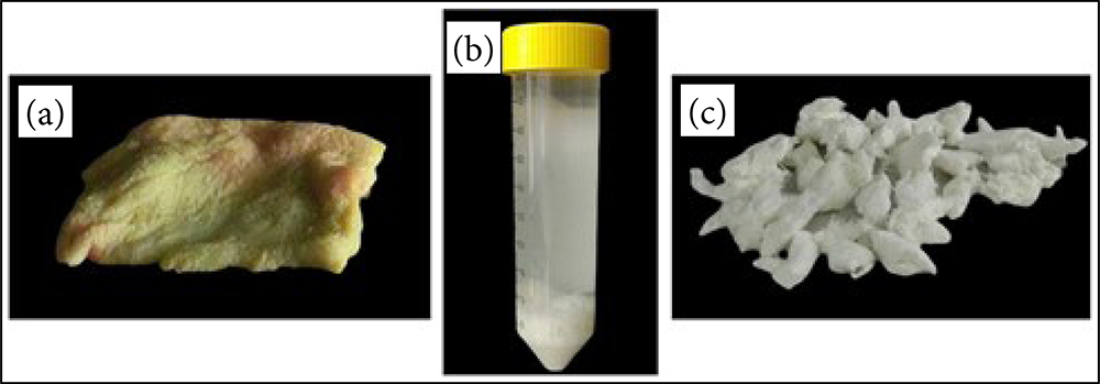
Figure 1. Porcine skin decellularization. (a) Porcine skin in natura. (b) Porcine skin triturated and incubated with reagents (trisol) during the decellularization process. (c) Decellularized and lyophilized extracellular matrix (n = 5).
Research carried out at the laboratories of the Faculty of Medicine of the University of São Paulo (FMUSP) analyzed a modified hydrogel production protocol to quantify the cellular content and toxicity of a matrix derived from decellularized swine skin, intended to create a scaffold for the proliferation of fibroblasts. The article A modified hydrogel production protocol to decrease cellular content published in vol. 37, no. 10 of the Acta Cirúrgica Brasileira journal presents the details of materials and procedures contemplated in the process, from the collection of the skin sample until the stages of decellularization, introduction and development of the fibroblast cell culture.
There were notable differences between the results obtained with the decellularized matrix hydrogel and that produced with in natura porcine skin composing, respectively, the experimental group and the control group. The two groups of samples were submitted to a histological analysis and quantification of the DNA present in the extracellular matrix. There was a comparison between the distribution of collagen fibers and the morphology of each group. The article brings the results of flow cytometry with the immunophenotyping of fibroblasts arranged in a histogram, while the cell culture in the hydrogel is illustrated in rendered images.
Cytotoxicity assay indicated that cell viability depends on hydrogel concentration. The adequate percentage found in the present study was observed between 3% and 25%. The conclusion of the article is that the analyzed process was effective in cell lysis and the presence of DNA elements was reduced to the impossibility of immunological rejection, but the decellularization process still needs to be improved to eliminate the cytotoxicity caused by detergents.
References
GILPIN, A. and YANG, Y. Decellularization strategies for regenerative medicine: from processing techniques to applications. Biomed Res Int [online]. 2017, vol. 2017, 9831534 [viewed 24 January 2023]. https://doi.org/10.1155/2017/9831534. Available from: https://www.hindawi.com/journals/bmri/2017/9831534/
ZHONG, S.P., ZHANG, Y.Z., LIM, C.T. Tissue scaffolds for skin wound healing and dermal reconstruction. Wiley Interdiscip Rev Nanomed Nanobiotechnol [online]. 2010, vol. 2, no. 5, pp. 510-525 [viewed 24 January 2023]. https://doi.org/10.1002/wnan.100. Available from: https://wires.onlinelibrary.wiley.com/doi/10.1002/wnan.100
To read the article, access
BRAGA, G.C.D., et al. A modified hydrogel production protocol to decrease cellular content. Acta Bras Cir [online]. 2022, vol. 37, no. 10, e371005 [viewed 24 January 2023]. https://doi.org/10.1590/acb371005. Available from: https://www.scielo.br/j/acb/a/g3wHWDMkqjq7HMJfPp39wCB/
External links
Acta Cirurgica Brasileira – ABC: https://www.scielo.br/acb/
Faculdade de Medicina da Universidade de São Paulo (FMUSP): https://www.fm.usp.br/
Acta Cirúrgica Brasileira – Social Media: Facebook | Twitter | Instagram
Como citar este post [ISO 690/2010]:


Recent Comments